Knee Pain Treatment

REQUEST A CALL BACK NOW!

What is a Knee Pain?

Knee pain is a common complaint that affects people of all ages. Knee pain may be the result of an injury, such as a ruptured ligament or torn cartilage. Medical conditions - including arthritis, gout and infections - also can cause knee pain. Many types of minor knee pain respond well to self-care measures. Physical therapy and knee braces also can help relieve knee pain. In some cases, however, your knee may require surgical repair.

Knee pain Knee pain

Symptoms

The location and severity of knee pain may vary, depending on the cause of the problem. Signs and symptoms that sometimes accompany knee pain include:

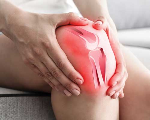
  • Swelling and stiffness
  • Redness and warmth to the touch
  • Weakness or instability
  • Popping or crunching noises
  • Inability to fully straighten the knee
  • Rolling the ankle

Knee pain can be caused by injuries, mechanical problems, types of arthritis and other problems.

Injuries

A knee injury can affect any of the ligaments, tendons or fluid-filled sacs (bursae) that surround your knee joint as well as the bones, cartilage and ligaments that form the joint itself. Some of the more common knee injuries include:
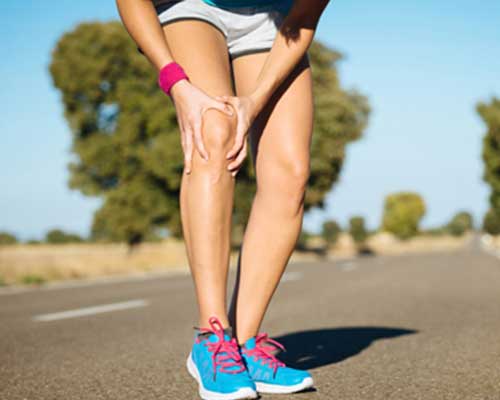
ACL injury.

An ACL injury is the tearing of the anterior cruciate ligament (ACL) - one of four ligaments that connect your shinbone to your thighbone. An ACL injury is particularly common in people who play basketball, soccer or other sports that require sudden changes in direction.

Torn meniscus.

The meniscus is formed of tough, rubbery cartilage and acts as a shock absorber between your shinbone and thighbone. It can be torn if you suddenly twist your knee while bearing weight on it.

Knee bursitis.

Some knee injuries cause inflammation in the bursae, the small sacs of fluid that cushion the outside of your knee joint so that tendons and ligaments glide smoothly over the joint.

Patellar tendinitis.

Tendinitis is irritation and inflammation of one or more tendons - the thick, fibrous tissues that attach muscles to bones. Runners, skiers, cyclists, and those involved in jumping sports and activities are prone to develop inflammation in the patellar tendon, which connects the quadriceps muscle on the front of the thigh to the shinbone.

Book An Appointment Here

Book An Appointment

Call Now!

Call Now

Mechanical Problems

Some examples of mechanical problems that can cause knee pain include:

  • Loose body. Sometimes injury or degeneration of bone or cartilage can cause a piece of bone or cartilage to break off and float in the joint space. This may not create any problems unless the loose body interferes with knee joint movement, in which case the effect is something like a pencil caught in a door hinge.
  • Iliotibial band syndrome. This occurs when the ligament that extends from the outside of your pelvic bone to the outside of your tibia (iliotibial band) becomes so tight that it rubs against the outer portion of your femur. Distance runners are especially susceptible to iliotibial band syndrome.
  • Dislocated kneecap. This occurs when the triangular bone (patella) that covers the front of your knee slips out of place, usually to the outside of your knee. In some cases, the kneecap may stay displaced and you'll be able to see the dislocation.
  • Hip or foot pain. If you have hip or foot pain, you may change the way you walk to spare these painful joints. But this altered gait can place more stress on your knee joint. In some cases, problems in the hip or foot can refer pain to the knee.

diagnosis

During the physical exam, your doctor is likely to:

  • Inspect your knee for swelling, pain, tenderness, warmth and visible bruising
  • Check to see how far you can move your lower leg in different directions
  • Push on or pull the joint to evaluate the integrity of the structures in your knee

Imaging tests

In some cases, your doctor might suggest tests such as:

  • X-ray. Your doctor may first recommend having an X-ray, which can help detect bone fractures and degenerative joint disease.
  • Computerized tomography (CT) scans. CT scanners combine X-rays taken from many different angles, to create cross-sectional images of the inside of your body. CT scans can help diagnose bone problems and detect loose bodies.
  • Ultrasound. This technology uses sound waves to produce real-time images of the soft tissue structures within and around your knee, and how they are working. Your doctor may want to maneuver your knee into different positions during the ultrasound, to check for specific problems.
  • Magnetic resonance imaging. MRI uses radio waves and a powerful magnet to create 3-D images of the inside of your knee. This test is particularly useful in revealing injuries to soft tissues such as ligaments, tendons, cartilage and muscles.

Lab tests

If your doctor suspects an infection, gout or pseudogout, you're likely to have blood tests and sometimes arthrocentesis, a procedure in which a small amount of fluid is removed from within your knee joint with a needle and sent to a laboratory for analysis.

Treatment

Treatments will vary, depending upon what exactly is causing your knee pain.

Medications

Your doctor may prescribe medications to help relieve pain and to treat underlying conditions, such as rheumatoid arthritis or gout.

THERAPY

Strengthening the muscles around your knee will make it more stable. Training is likely to focus on the muscles on the front of your thigh (quadriceps) and the muscles in the back of your thigh (hamstrings). Exercises to improve your balance are also important.

Arch supports, sometimes with wedges on one side of the heel, can help to shift pressure away from the side of the knee most affected by osteoarthritis. In certain conditions, different types of braces may be used to help protect and support the knee joint.

ligament ligament ligament ligament

Copyright © 2018 Chaubey's Physio All rights reserved. Design & Develop by WebzSpan Technologies